Computed Ultrasound (CUS)
MRI-level 3D imaging in 3 minutes
AI-powered handheld imaging that delivers submillimeter 3D diagnostic scans — operable by any healthcare worker, anywhere in the world.
- Handheld probe, no specialist equipment needed
- Full 3D volume output in standard NIfTI format
- Any HCA can operate it — no radiographer required
- UK MDR 2002 compliant — no MHRA device registration required
- Works in clinics, rural settings, and field hospitals
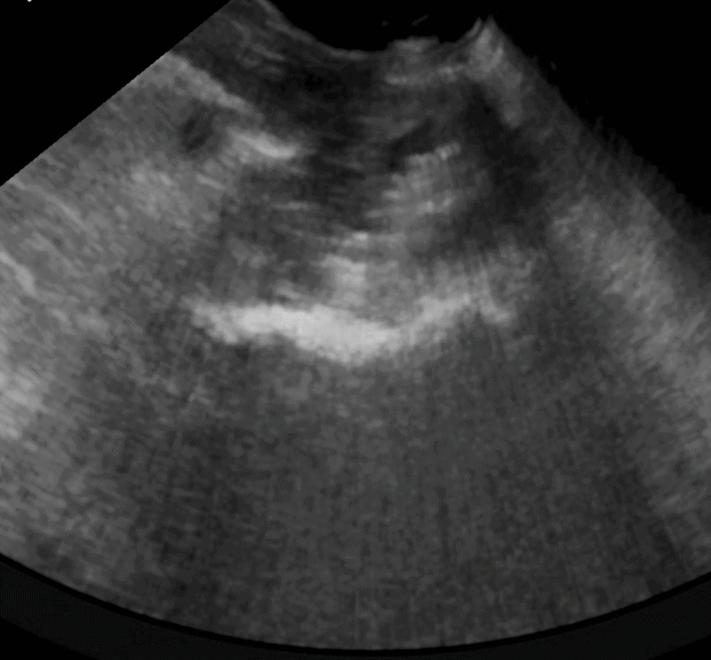
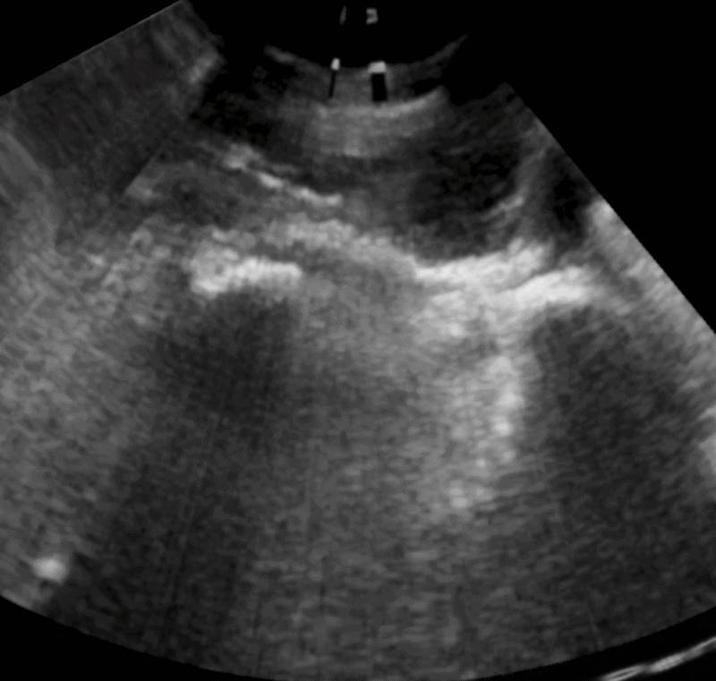
Cardiac Module
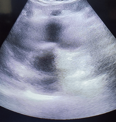
Live 3D CUS scan
<3 min
Full 3D scan time vs 30–45 min for MRI
<1mm
Voxel resolution — comparable to MRI
94%
Gross margin on a pure SaaS model